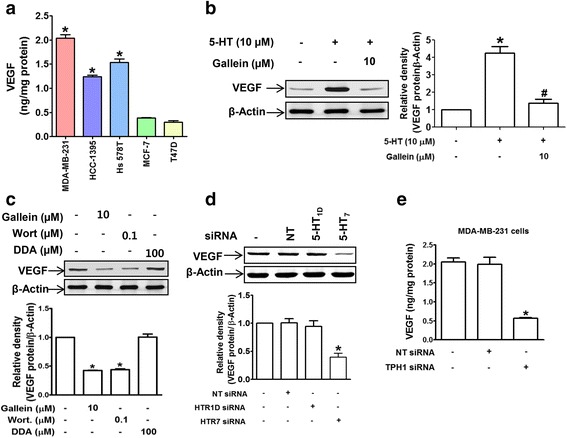
Fig. 4.

5-HT crosstalks with VEGF by inducing its expression. a VEGF secretions by TNBCs and hormone-responsive breast cancer cells were measured by ELISA. *P < 0.05 vs. T47D cells. b Cells were treated as described in the legend of Fig. 3c and VEGF protein levels were determined by western blotting. *P < 0.05 vs. vehicle-treated controls. # P < 0.05 vs. 5-HT-treated cells. c Basal levels of VEGF protein in MDA-MB-231 cells treated with gallein, wortmannin, or DDA. The bar graphs represent the means ± SEMs of three independent experiments. *P < 0.05 vs. vehicle-treated controls. d Knockdown of 5-HT7 suppressed VEGF protein expression. *P < 0.05 vs. vehicle- or NT siRNA-treated controls. e Knock-down of TPH1 inhibited VEGF secretion by MDA-MB-231 cells. *P < 0.05 vs. vehicle- or NT siRNA-transfected controls
