Fig. 3.

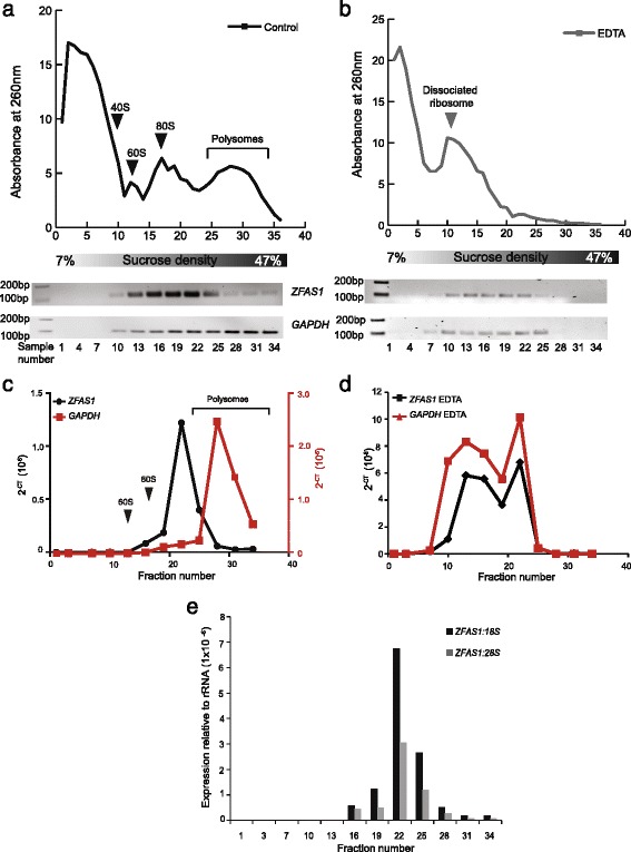
ZFAS1 is associated with actively translating ribosomes. a Polysome distribution of MDA-MB-468 cell lysates as separated on a 7–47% sucrose gradient. Absorbance at 260 nm is shown on the Y axis. Fractions from the top of the gradient to the bottom are shown from left to right on the X axis. Fractions were collected in 36 equal volumes, of which every third was used for RNA extraction, and cDNA synthesised for PCR. The presence of ZFAS1 expression was assessed using primers described in Fig. 1, with GAPDH acting as a positive control. Both species were enriched in the bottom fractions of the gradient, corresponding to polysomes. b Polysome distribution of MDA-MB-468 cell lysate separated on a 7–47% sucrose gradient containing EDTA instead of MgCl2. Loss of the polysome peak is observed, together with a leftward shift of the ribosome subunits. RT-PCR analysis (lower panels) showed that ZFAS1 and GAPDH are no longer enriched in the lower peaks, and show concomitant shifts to the upper fractions. c and d Quantitative expression of ZFAS1 and GAPDH measured by qPCR relative to 18S and 28S rRNAs prepared with and without the addition of EDTA (black graph for ZFAS1, red graph for GAPDH). ZFAS1 and GAPDH are no longer enriched in the lower fractions corresponding to polysomes following polysome disruption by EDTA. Arrows indicate where ribosomal features are observed on profiles in relation to fraction number. e Ratio of ZFAS1 to 18S and 28S rRNA in fractions from MDA-MB-468 polysome gradients. qPCR was performed using samples from polysome separation and expression of ZFAS1 relative to 18S and 28S was calculated. ZFAS1 was detected in fractions 16–34 and showed greatest abundance in fraction 22, corresponding to light polysomes. The ratio of ZFAS1: 18S is also greater than that of ZFAS1:28S for all fractions
