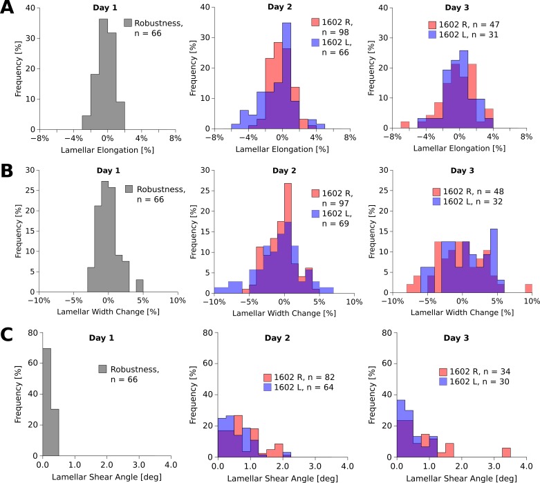
Fig 10.
Histograms of lamellar elongation (A), width change (B), and shear angle distributions (C) of the left (L) and right (R) eye of tree shrew 1602 over three days in organ culture. The lamellar elongation (A) and width changes (B) were normally distributed around zero at all days of organ culture. The variance of the distributions significantly increased in organ culture (Levene’s test, P < 0.01). The shear angle (C) significantly increased in both eyes (Kruskal-Wallis test, P < 0.01). Robustness values were measured after the eye chamber was tilted on day 1 of organ culture.

