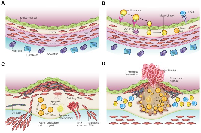
Figure 4.
Schematic overview of the development of an atherosclerotic lesion. In all steps, inflammation plays an important role. (A) A healthy artery with a well-functioning intact endothelium, a tunica intima, media and adventitia. VSMCs are mainly found in the tunica media but also in the tunica intima. (B) One of the initiating steps is the expression of adhesion molecules on the endothelium and the subsequent attraction of inflammatory blood cells (mainly monocytes). These monocytes will transmigrate to the intima where they will maturate to macrophages which will then transform to foam cells upon the uptake of ox-LDL. (C) Further progression to an atherosclerotic plaque includes the transmigration of VSMCs from the tunica media into the intima and the proliferation of VSMCs in the intima. There is also an enhanced production of extracellular matrix molecules, such as collagen, elastin and proteoglycans. Macrophages, foam cells and VSMCs can die, and released lipids will accumulate into the central region of the plaque, also denoted the lipid or necrotic core. (D) When a plaque ruptures it will induce thrombosis which is the major complication. The blood component will come in contact with the tissue factors present in the interior of the plaque triggering the formation of a thrombus which will hamper or even obstruct blood flow. The figure is based on a previous study (167). VSMCs, vascular smooth muscle cells; ox-LDL, oxidized low-density lipoprotein.

