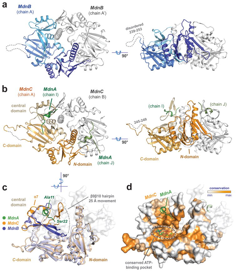
Figure 3. Overall structures of MdnC and MdnB.
(a) Two views of MdnB (blue/grey) and (b) MdnC (orange/grey) dimers. MdnC forms a co-complex with the precursor peptide MdnA1–35 (green). (c) Superimposition of the MdnC and MdnB protomers illustrate domain movement involved in precursor peptide binding. (d) Evolutionary conservation map of MdnC protomer highlights conserved regions of the C-domain. The modelled ADP/Mg2+ binding site is noted on the map.

