Figure 2.
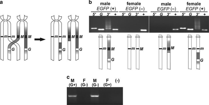
Loss of linkage between the maleness locus and the EGFP tag during male meiosis. (a) A diagram of a crossover between two non-sister chromatids during meiotic prophase, giving rise to recombinant chromosomes (only pericentromeric fragments of chromosomes 1 are presented). The sex locus (black line or square denoted either m or M) and the transgene (white square denoted G) are shown. (b) A PCR confirming integrity of the junctions in the EGFP-positive individuals and indicating loss of the transposon in males and gain thereof in females in a generation following the recombination. The phenotypes of individuals analyzed are given at the top. For each phenotype, lanes are marked as follows: 5′, a product spanning the 5′ junction; G, a fragment of the EGFP gene; 3′, a product spanning the 3′ junction; +, a fragment of the Aams gene (positive control of DNA quality). A smeary ladder-like pattern of the PCR product spanning the 3′ junction results from binding of the near_pBac3′_2 primer to multiple target sites within a tandemly repeated flanking DNA. Combinations of chromosome 1 pairs corresponding to each phenotype are depicted below the gel image. (c) Test for presence of myo-sex in the EGFP-positive and EGFP-negative individuals, denoted as G+ or G−. M, male; F, female; (−), negative control.

