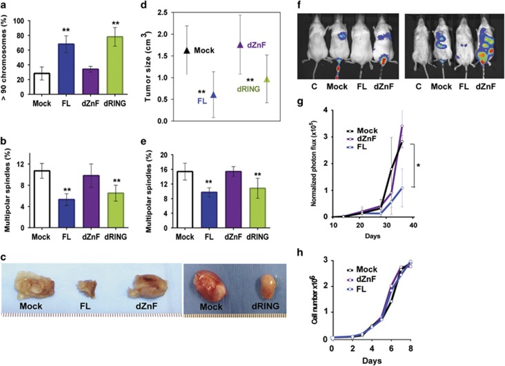
Figure 5.
The Zn-finger domain of MdmX suppresses tumor growth and metastatic potential of human tumor cells. (a) Chromosome analyses showing percent of cells with >90 chromosomes per cell in populations of human MB231 breast tumor cells transduced with MdmX FL or deletion mutants. Error bars represent mean±s.d. from four to six mitotic spreads with at least 30 metaphase cells scored per cell line per spread. (b) Frequency of multipolar spindles in populations of transduced MB231 cells. More than 200 mitotic events were scored per cell line per experiment. Error bars represent mean±s.d. from four experiments. (c) Tumors formed after orthotopic transplantation of MB231 cells into the mammary fat pad of nude mice. Tumors were harvested 16 days after transplantation. (d) Tumorigenic potential of transduced MB231 cells transplanted into the fat pad of nude mice. Tumor size was calculated from equation: cm3=½ (W2 × L). The error bars represent mean±s.d. of tumor size from 3 to 5 animals per each cell line. (e) Frequency of multipolar spindles in populations of cells cultured from tumors harvested after transplantation of transduced MB231 cells into the mammary fat pad of nude mice. More than 500 mitotic events were scored in population of cells deriving from each tumor. Error bars represent mean±s.d. from three to five tumors. (f) Representative bioluminescence images of lung colonization in mice. MdmX-transduced MB231 cells infected with retroviral M-cherry-Luciferase reporter were injected into the tail vein of nude mice. Lung colonization was assayed by bioluminescence imaging following intraperitoneal injection of Luciferin. From left to right: control for auto-luminescence (cells without retrovirus); Mock (cells without MdmX); cells with FL MdmX; cells with MdmX-dZnF mutant. (g) Metastatic potential of transduced MB231 cells expressing Luciferase reporter, injected into the tail vein of nude mice (2.5 × 10⁵ cells per animal, five animals per group). Luciferase activity was monitored over time using IVIS 100 imager. Intensity of photon flux within the region of interest from combined dorsal and ventral images of animals within the group was normalized to the intensity of photon flux on day 1. (h) Proliferation of MdmX-transduced MB321 cells used in experiments presented in a–g. P-values relative to Mock control: **P<0.005; *P value from 0.05 to 0.005, unpaired t-test.

