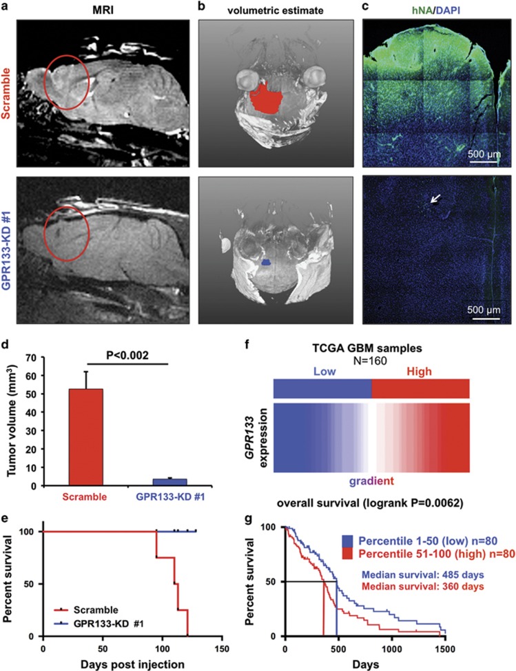
Figure 6.
GPR133 knockdown prevents tumor formation and death in vivo. (a and b) Representative MRI images (a) and tumor volumetric estimates (b) show marked reduction in tumor xenograft size in mice implanted with GBM cells bearing GPR133 shRNA knockdown construct no. 1 (GPR133-KD no. 1) (GBML20, n=4 animals per group). (c) Histology of tumor xenografts. Tumor xenografts were identified by human nuclear antigen (hNA) immunoreactivity. The arrow indicates scattered tumor cells in the GPR133-KD condition, in the absence of formed tumor. (d) Cumulative statistics for tumor size obtained from MRI-based volumetric estimates (GBML20, P<0.002, t-test, n=4 animals per group). (e) Kaplan–Meier survival curves of the GPR133-KD no. 1 and control groups (log-rank Mantel–Cox test, P=0.0143). (f) The TCGA data from 160 patients with GBM were analyzed for GPR133 expression. We studied outcomes in two cohorts GPR133 high (in red) and GPR133 low (in blue) based on ranked GPR133 mRNA levels. (g) Kaplan–Meier curves of the two patient cohorts indicate an inverse relation between GPR133 expression and survival (log-rank Mantel–Cox test, P=0.0062).

