Figure 4.
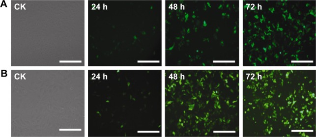
GFP expression in transfected HepG2 cells.
Notes: CK: untransfected cells; 24, 48, and 72 h represent different time points post-transfection. Cells transfected GFP with PEI/QDs (A); Cells transfected GFP with Lipo2000 (B). Scale bar =100 μm.
Abbreviations: GFP, green fluorescent protein; PEI, polyethyleneimine; QD, quantum dot.

