Figure 5.
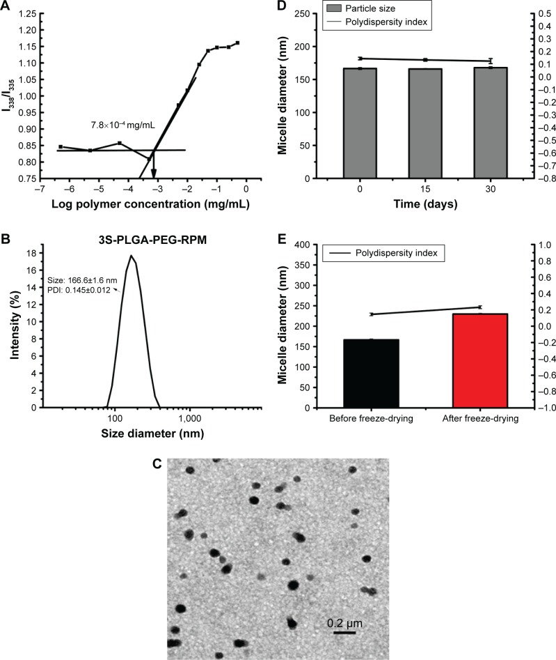
(A) CMC of 3S-PLGA-PEG: pyrene was used as the fluorescence probe in the measurement. With increasing polymer concentrations in aqueous solution, I338/I335 values in the excitation spectrum increased. The straight line below CMC represents the stable state of solution, the straight line above CMC represents the stable state of micellar solution, the point of intersection of these two lines are the Krafft point, which represents the CMC. The CMC of 3S-PLGA-PEG was 7.8×10⁻⁴ mg/mL, which suggested that the micelles formed easily. (B) 3S-PLGA-PEG-RPM micelles were small (166.6±1.6 nm, PDI 0.145±0.012), which suited their circulation in blood, and had passive targeting to tumor tissue. (C) TEM suggested that micelles obtained appeared similarly spherical in shape and separated from each other. The results of DTS and TEM indicated that 3S-PLGA-PEG micelles possessed ideal size and good dispersibility. (D) The stability of the micelle suspension at 1 month suggested that the 3S-PLGA-PEG micelles were very stable during storage in 4°C. (E) The slight difference between before freeze-drying and after freeze-drying indicated slight sedimentation and aggregation.
Abbreviations: CMC, critical micelle concentration; 3S-PLGA, three-arm star block poly(lactic-co-glycolic acid); PEG, polyethylene glycol; RPM, rapamycin; PDI, polydispersity index; TEM, transmission electron microscopy; DTS, dynamic light scattering.

