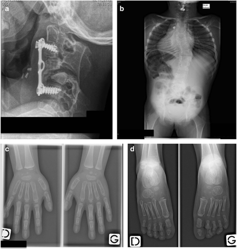
Figure 4.
Radiographic examination of patient 2-II.1 at 5 years and 6 months of age (a) and at 3 years and 8 months of age (b, c, d). (a) Lateral X-ray of cervical spine 3 months after a C3–C6 anterior epiphysiodesis with tibial bone graft and plate fixation showing an incomplete reduction of a C4–C5 dislocation and posterior spinal fusions. (b) PA X-ray of thoracolumbar spine showing thoracic vertebral fusions without rib fusions responsible for growth disturbance with a short trunk and a right thoracolumbar curve. (c) AP X-rays of hands showing capitate–hamate coalition in both hands. (d) Dorsal plantar X-rays of feet where no coalition is observed.

