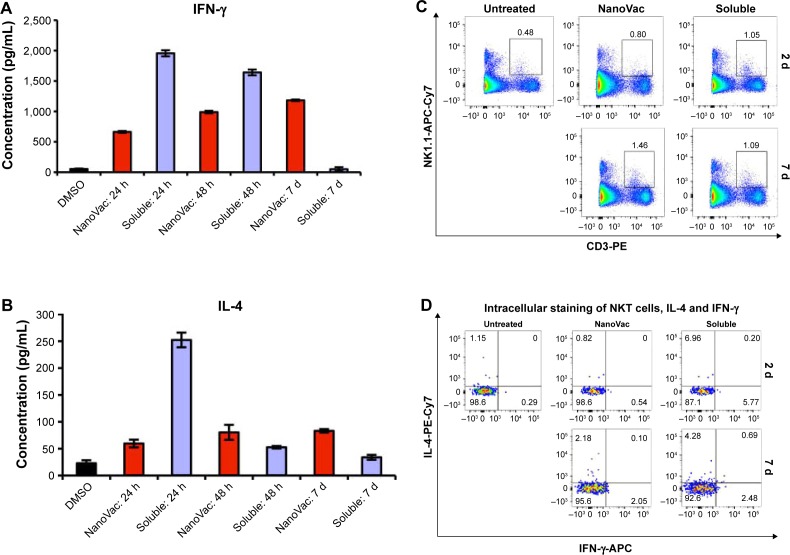
Figure 4.
Characterization of cytokine release and NKT cell proliferation after the treatment of nanovaccines.
Notes: The presented data are representative of three mice. (A and B) In vivo assessment of secreted IFN-γ and IL-4 from splenocytes after treatment with NanoVac and soluble Vac. Three time points were chosen as indicated. (C) Evaluation of NKT cell proliferation. Each group (n=6) of mice received treatment as indicated. Flow cytometry studies were performed to measure the proliferation of NKT cells on days 2 and 7. (D) Intracellular cytokine staining. The staining of IFN-γ and IL-4 was done in NKT cells from (C).
Abbreviations: APC, antigen-presenting cell; d, days; DMSO, dimethylsulfoxide; h, hours; IFN-γ, interferon-γ; IL, interleukin; NKT, natural killer T.

