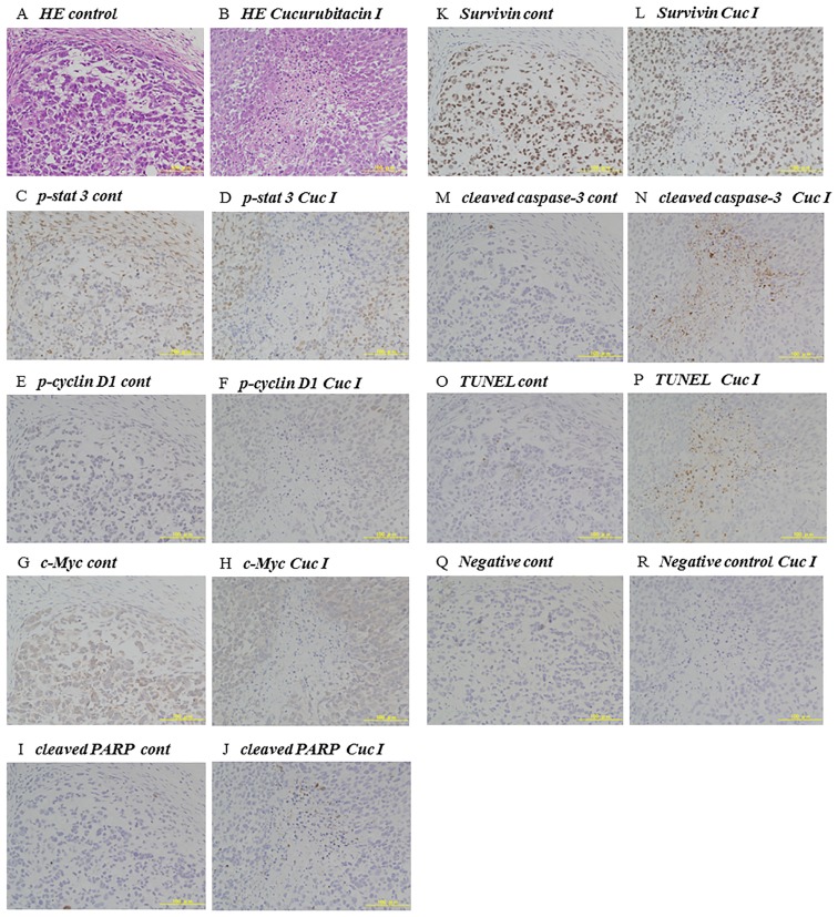
Figure 6.
Immunohistochemical analysis of cucurbitacin I-treated tumors. Representative 143B tumors of athymic nude mice were immunohistochemically analyzed on day 14 after administration of control (A, C, E, G, I, K, M, O and Q) or 1.0 mg/kg cucurbitacin I (B, D, F, H, J, L, N, P and R). (A and B) Hematoxylin-eosin (H&E) staining. (C and D) Immunohistochemical staining of phospho-STAT3. (E and F) p-cyclin D1. (G and H) c-Myc. (I and J) Cleaved PARP. (K and L) Suvivin. (M and N) Cleaved caspase-3. (O and P) TUNEL assay. (Q and R) Negative control.

