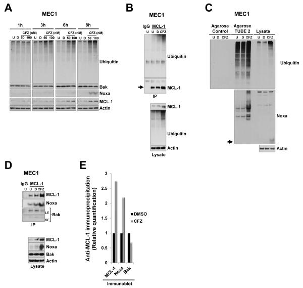
Figure 3. Carfilzomib treatment resulted in accumulation of polyubiquitinated MCL-1 and Noxa.
(A) Carfilzomib treatment results in accumulation of MCL-1 and Noxa protein in a time dependent manner. MEC1 cells were incubated for the indicated time with vehicle or with the indicated concentrations of carfilzomib or left untreated. Cell lysates were subjected to immunoblotting analysis with the indicated antibodies. (B) Accumulated MCL-1 proteins were polyubiquitinated in response to carfilzomib. MEC1 cells were left untreated or were treated with vehicle or 25 nM carfilzomib for 24 h. Cell lysates were immunoprecipitated (IP) with MCL-1 antibodies. To show specificity, IgG antibodies were used in parallel in an immunoprecipitation assay with untreated cell lysate. Bound proteins were subjected to SDS-PAGE and then immunoblotted with ubiquitin and subsequently with MCL-1 after stripping of the membrane (upper panels). Cell lysates were also immunoblotted with the indicated antibodies (bottom panels). Arrow indicates full-length form of the corresponding protein at the expected molecular weight. (C) Accumulated Noxa proteins were polyubiquitinated in response to carfilzomib. Agarose beads or agarose TUBE 2 beads were mixed with cell lysates from MEC1 treated as indicated in (B). Bound proteins were subjected to SDS-PAGE and immunoblotted with ubiquitin (upper panel) or Noxa (lower panel). Arrow indicates full-length form of the corresponding protein at the expected molecular weight. Respective cell lysates were run in parallel on the same gel (right portion of the blot). (D) Accumulated MCL-1 and Noxa preferentially formed a complex after carfilzomib treatment. Cell lysates from MEC1 cells that were treated as indicated in (B) were subjected to immunoprecipitation (IP) with MCL-1 antibodies (upper panels). Bound proteins were subjected to SDS-PAGE and immunoblotted with Noxa and Bak antibodies and then with anti-MCL1 after stripping. To show specificity, IgG antibodies were used in parallel in an immunoprecipitation assay with untreated cell lysate. In parallel, respective cell lysates were also subjected to immunoblot analysis with the indicated antibodies (lower panels). U, untreated; D, DMSO; CFZ, carfilzomib; LE, long exposure; SE, short exposure. (E) MCL-1/Noxa complexes were prevalent from MCL-1/Bak complexes after carfilzomib treatment. Densitometry quantification of the immunoprecipitation shows in (D).

