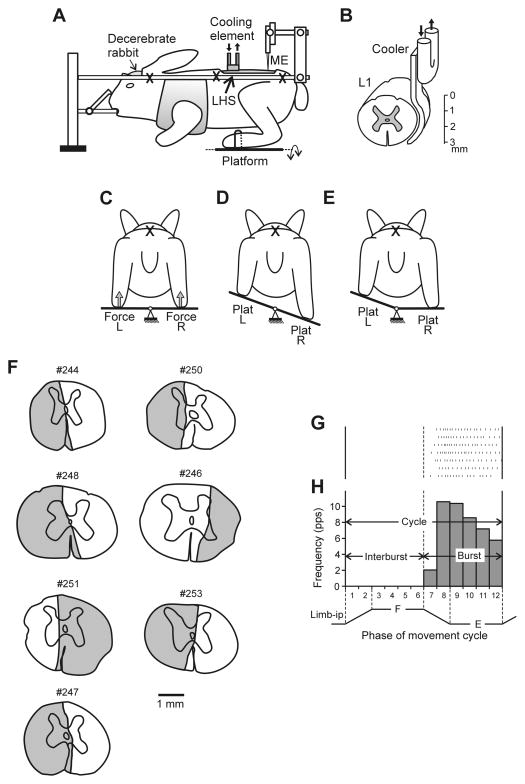
Fig. 1. Experimental design.
(A) the decerebrate rabbit was fixed in a rigid frame (points of fixation are indicated by X). In a part of experiments surgical lateral hemisection of the spinal cord (LHS) was performed at L1 (indicated by red arrow). In a part of experiments the cooling element was positioned on the lateral surface of the spinal cord at L1. (B) Position of the cooler on the lateral surface of the spinal cord. To evoke postural limb reflexes (PLRs), the hind limbs were positioned on a platform (C), which was periodically tilted in the transversal plane either as a whole (D), or its left (Plat L) and right (Plat R) parts were tilted separately (E). The contact forces under the left and right limbs were measured by the force sensors (Force L and Force R in C). Activity of spinal neurons from L5 was recorded by means of the microelectrode (ME in A). (F) Extent of the spinal cord damage in rabbits with lateral lesions. The total extent of the lesion is projected on a spinal cord section taken more rostrally after inspecting several consecutive sections. (G) A raster of responses of E-neuron in 7 sequential movement cycles of the ipsilateral limb. (H) A histogram of spike activity in different phases (1–12) of the cycle of movement (F, flexion; E, extension) of the ipsilateral limb (Limb-ip). The halves of the cycle with higher (E, bins 7–12) and lower (F, bins 1–6) neuronal activity were designated as “Burst” and “Interburst” periods, respectively. A,C–D is modified from Fig. 2A–D in Musienko et al., 2010.

