Figure 3.
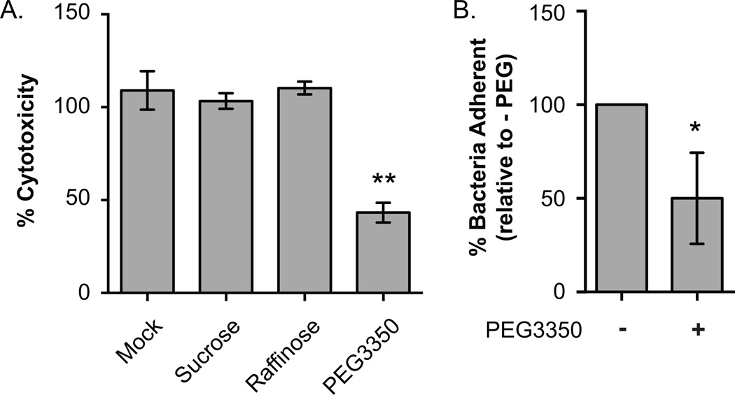
(A) Pore formation influences cytotoxicity. Three independent colonies of the AM-19226 T3SS WT strain were grown overnight in LB and used to infect Caco2-BBE cells at an MOI of ~10 in the presence of 0.2% bile with or without 30mM sucrose, 30mM raffinose, or 30mM PEG3350. Percent cytotoxicity was calculated at 3h after co-culture began by measuring LDH released into the supernatant. Data represent the mean ± standard deviation from one representative experiment. The experiment was repeated once with similar results. **, P<0.0001 by one-way ANOVA with Dunnett post hoc test. (B) Bacterial adherence in the presence of PEG. AM-19226 T3SS WT was grown overnight in LB and co-cultured with Caco2-BBE and an MOI of ~10 in the presence of 0.2% bile with or without 30mM PEG3350. After 2h, percent of bacteria adherent to Caco2-BBE cells was determined. Data represent the mean ± standard deviation from three independent experiments using four individual bacterial colonies per experiment. * P<0.05 by t-test.

