
Fig. 9.
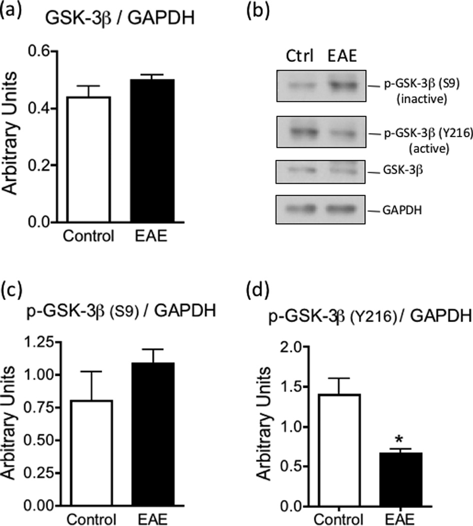
GSK-3β activity is reduced in EAE. The levels of GSK-3β (panel a), phospho-GSK-3β (S9) (panel c) and phospho-GSK-3β (Y216) (panel d) in the spinal cord of control and EAE mice at 21 dpi were determined by western blot analysis as described under “Materials and Methods” and were normalized by the amount of GAPDH same gel lane. Values represent the mean ± SEM of 3–4 animals per experimental group. Asterisk denotes the value that is statistically different (p<0.05) from the control. Panel b shows representative western blots.