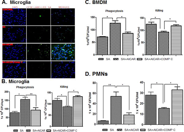
Figure 8. AICAR treatment increases the phagocytic activity of microglia, macrophages, and neutrophils.

CFSE-labelled S. aureus was used to challenge the BV2 cells (5 × 10⁵ cells/chamber) (n=4/condition) cultured in chamber slides under the indicated conditions (i.e., AICAR or Compound C treatment) for 2h. Stained cells were subjected to fluorescent imaging to visualize intracellular S. aureus (A). In another experiment, to assess phagocytic activity, cultured BV2 microglial cells) (B), bone-marrow derived macrophages (C) and neutrophils (D) were left untreated or treated with AICAR (1mM) and Comp C for one hour prior to S. aureus challenge (n=4/condition), as described in ‘Materials and Methods’. After 2 h of bacterial challenge, the cells were washed and kept in fresh medium containing gentamicin (200 μg/ml). After 2h of incubation, the cells were washed and lysed by using Triton-× (0.1%). The release of intracellular bacteria was quantitated via serial dilution and plate count (B, C & D). In the killing assay, cells were incubated with S. aureus for 2h, after which Triton × (0.1%) was added to the cells. Cells were then serially diluted and plated for counting (B, C & D). The data points represent the mean ± SD of three independent experiments. Statistical analysis was performed using a one-way ANOVA with Bonferroni’s multiple-comparison test. * p < 0.05, ** p < 0.001.
