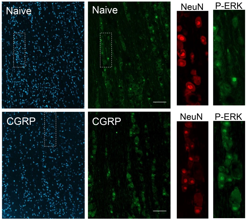
Figure 4.
Intracisternal CGRP injection increased expression of P-ERK in trigeminal ganglion neurons. Representative images of sections from the V1/V2 region of trigeminal ganglia obtained from naïve and CGRP treated animals are shown. All cell nuclei are identified by the nuclear dye DAPI (left panel), while the same tissue sections that were positive for P-ERK are seen in the second panel. Enlarged images of the region of the ganglion containing numerous neuronal cell bodies (white box) stained for neuronal protein NeuN (third panel) and the same region co-stained for P-ERK (far right panel) are shown. Scale bars = 100 μm.

