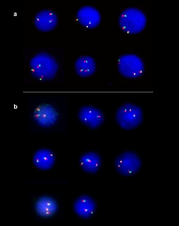
Figure 1.

(A) Dual‐probe FISH analysis using two probes for different loci on chromosome 21 showing three green and three red signals in cells from a pregnant women carrying a trisomy 21 fetus (patient B42). Yellow signals results from overlapping of green and red signals. (B) Dual‐probe FISH analysis using two probes for different loci on chromosome 18 showing three signals in green and three signals in red in cells from a pregnant women with a trisomy 18 fetus (patient C46). Yellow signals results from overlapping of green and red signals.
