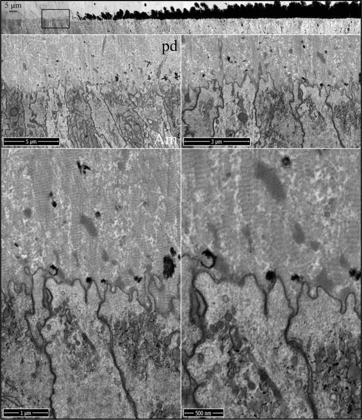
Figure 3.

Focused ion beam images of the onset of dentin mineralization near ameloblasts in an Enam ‐/‐ mouse mandibular incisor. Top: Low magnification montage of an incisor cross sectioned at Level 1 (~1 mm from its basal end). The box shows the region detailed by higher magnification images. Banded collagen fibers butt into ameloblasts at nearly right angles. Enamel matrix is accumulating in predentin. Key: Am, ameloblast; pd, predentin.
