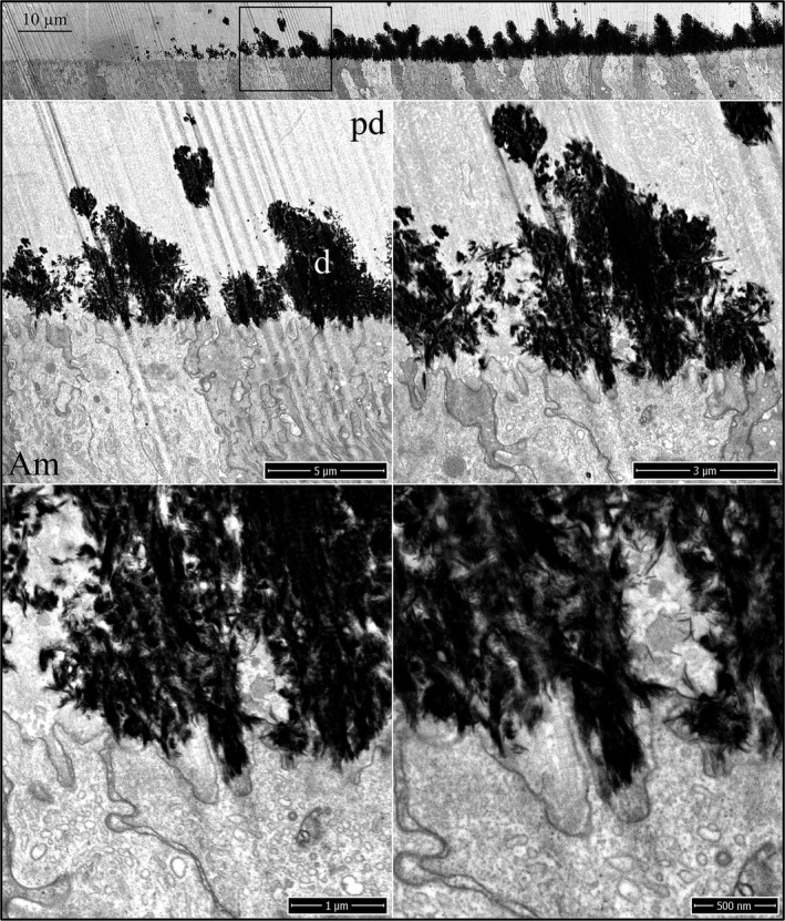
Figure 5.

Focused ion beam images of dentin mineralization near ameloblasts in an Amelx ‐/‐ mouse mandibular incisor. Top: Low magnification montage of incisor region as characterized at Level 1. The box outlines the region detailed by higher magnification images shown below. Prior to the coalescing of dentin mineral into a continuous layer along the irregular ameloblast surface, enamel mineral ribbon formation has not yet initiated. Key: Am, ameloblast; d, dentin; pd, predentin.
