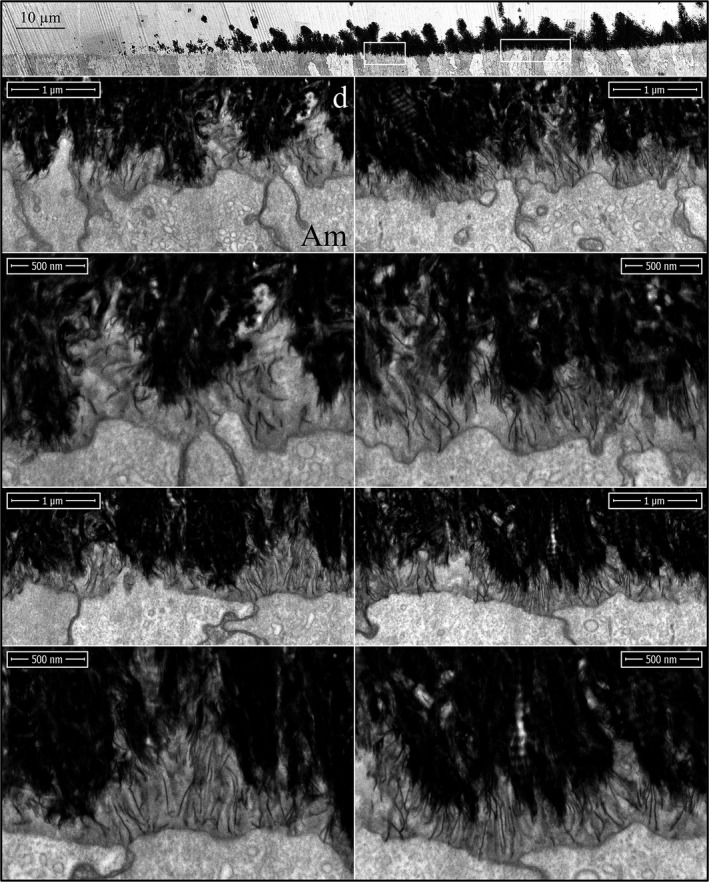
Figure 8.

Focused ion beam images of the onset of enamel mineralization in a Amelx ‐/‐ mouse mandibular incisor. The first enamel ribbons form on collagen mineral near the ameloblast membrane and orient in the path that the ameloblast process that initiated retreated into the distal membrane. The initial ribbons are short and elongate much more slowly than the wild‐type. The ameloblast distal membranes has fewer invaginations. Key: Am, ameloblast; d, dentin.
