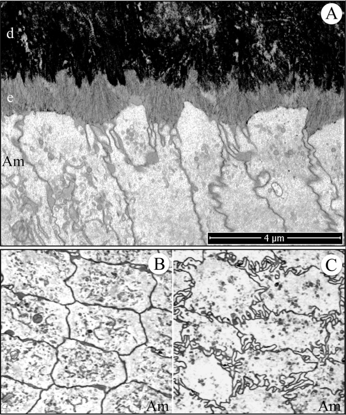
Figure 11.

Focused ion beam image of initial enamel formation in a wild‐type mouse mandibular incisor. (A) Longitudinal image from the serial set used for tomographic reconstruction. Note that the ameloblast distal membrane is more invaginated near the intercellular junctions and that clusters of enamel ribbons travel at different angles from the dentin to the ameloblast. This figure shows the scale for the Videos S22 and S23. (B, C) Cross‐sectional images captured from the tomographic reconstruction videos showing the relatively smooth ameloblast membrane proximal to the highly convoluted ameloblast membrane near the mineralization front. Key: Am, ameloblast; d, dentin; e, enamel.
