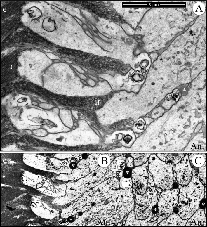
Figure 13.

Focused ion beam image of secretory stage enamel formation in a wild‐type mouse mandibular incisor. (A) Image from the serial set used for the making the tomographic reconstruction videos (Videos S27 and S28) and provides a scale bar for them. (B) Longitudinal section captured from the tomographic video (Video S27). (C) Cross section captured from the tomographic video (Video S28). Note the dense, droplet‐like accumulations of secreted proteins proximal to the distal cell junctions. Key: Am, ameloblast; asterisk, interproximal matrix accumulation; e, enamel; r, rod enamel; ir, interrod enamel.
