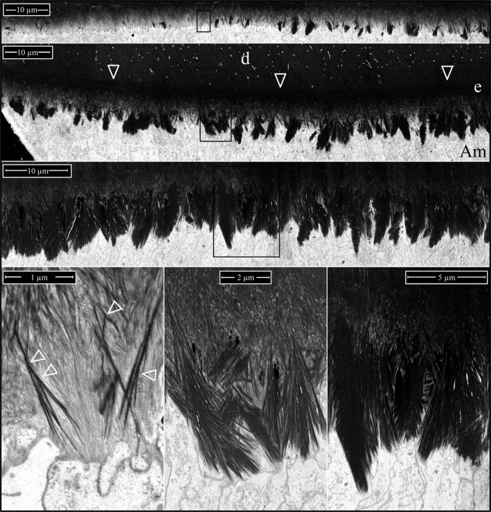
Figure 15.

Focused ion beam images of Amelx ‐/‐ Level 2 enamel (osmicated). The top three panels are montages of the Level 2 section on the lateral, midlateral, and central aspects of the incisor. Arrowheads point to the DEJ. Boxes delineate the three regions detailed by the higher magnification images are shown below (left to right, respectively). Arrowheads indicate sites of apparent crystal fusions. Key: Am, ameloblast; d, dentin; e, enamel.
