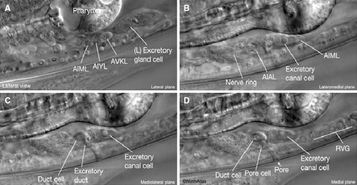
Fig. 11.
Differential interference contrast microscopy image of the excretory system in an adult C. elegans. a The excretory gland cell is located on the ventral side between the intestine and the terminal bulb of the pharynx. b The excretory cell nucleus is large and has a “fried egg” appearance with a large nucleolus. c The duct cell is anterior to the excretory cell d The excretory pore cell is ventral to the duct cell. The duct passes through the duct and pore cells and opens outside at the pore. RVG; retrovesicular ganglion (Altun and Hall 2012)

