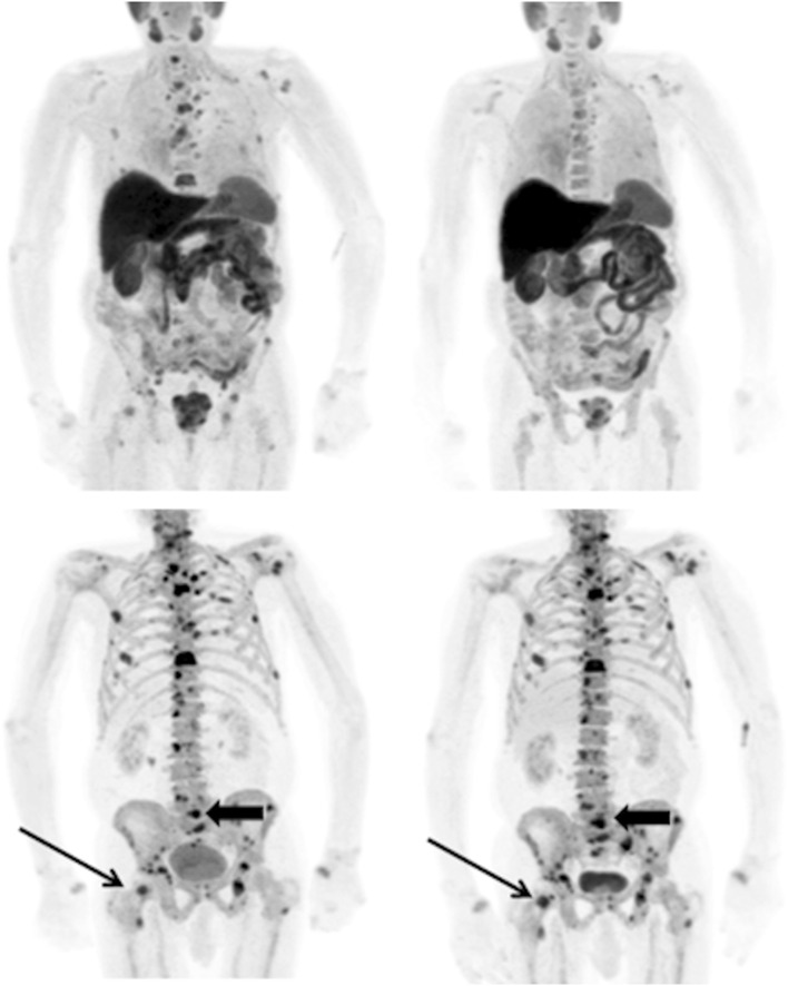
Fig. 1.
A patient with metastatic prostate cancer undergoing treatment with docetaxel chemotherapy. Top row 11C-choline PET maximum intensity projection images at baseline (left) and 8 weeks (right) and bottom row corresponding 18F-fluoride PET images. The higher contrast between metastases and the normal skeleton on the 18F-fluoride scans compared to the 11C-choline scans allows easier detection of disease. However, whilst there is a clear metabolic response in the bone metastases on the 11C-choline scans, there is a similar distribution and intensity of most lesions on the 18F-fluoride scans and some lesions show an increase in activity (arrows). This is likely to be due to a flare response at 8 weeks on the 18F-fluoride PET scans limiting the sensitivity and specificity in response prediction at early time points with this tracer as changes in osteoblastic activity lag behind changes in tumour metabolism

