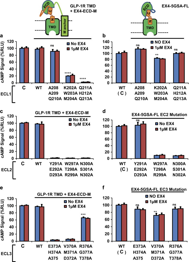
Figure 8.
The interaction interface between ECD and TMD localizes to the ECL of GLP-1R. Top left: cartoon representation of receptor TMD co-expressed with membrane-tethered EX4-ECD fusion (EX4-ECD-M); this construct has been used in a, c and e. Top right: cartoon representation of the hormone-fused receptor; this construct has been used in b, d and f. (a, c and e) cAMP signal produced by EX4-ECD-M and GLP-1R TMD with indicated mutations in ECL1, 2 and 3. (b, d and f) cAMP signal produced by EX4-5xGSA-GLP-1R FL construct harboring mutations in the same sites as the constructs in (a, c and e). All signal are indicated as relative to control (EX4-5xGSA-GLP-1R FL WT, labeled as C in a, c and e, as C with bracket in b, d and f), which was set to 100%. EX4-ECD-M: membrane-tethered EX4-ECD fusion. Error bars=s.d., all experiments were performed as three independent transfection experiments; two-tailed Student’s t test was used to determine P-values for data points versus WT: NS>0.05; *⩽0.05; **⩽0.01; ***⩽0.001; ****⩽0.0001. See Supplementary Figure S3B–D for activities normalized to expression levels.

