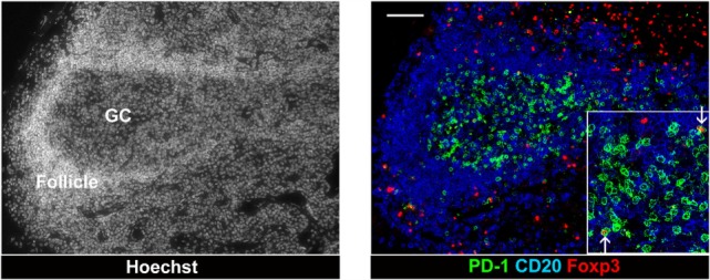
Figure 3.

Follicular T regulatory cells in a hyperplastic follicle of lymph node from chronic SIV infection. Foxp3+ cells and PD-1 high cells in an expanded GC of a hyperplastic follicle. The lymph node biopsies were stained with CD20 (blue), PD-1 (green), Foxp3 (red), and Hoechst dye (white). Scale bars = 50 μm.
