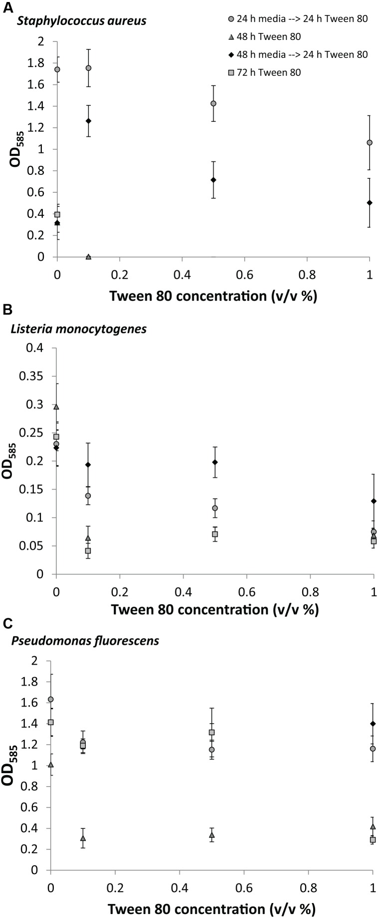
FIGURE 2.

Crystal violet staining of (A) S. aureus, (B) L. monocytogenes, (C) P. fluorescens after growth of biofilms for 0, 24, or 48 h in TSB followed by treatment with 0.1, 0.5, or 1.0% (v/v) Tween 80 for 24, 48, or 72 h. Error bars show standard deviation of the mean. All treatments were statistically significant (p < 0.05) from the control, except S. aureus 0.1% Tween (24 h→24 h); L. monocytogenes 0.1% and 0.5% Tween (48 h→24 h); and P. fluorescens 0.5% and 1% (48 h→24 h).
