Figure 6.
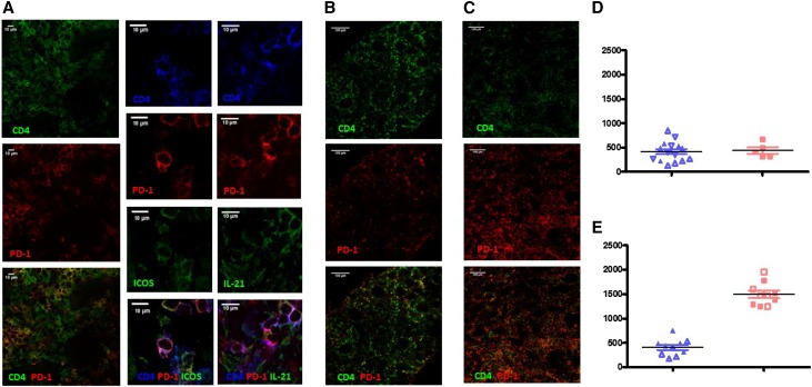
Tfh–like graft infiltrating lymphocytes were elevated in biopsies from belatacept-treated animals. (A) Immunostaining of a protocol biopsy showing cells with a Tfh-like phenotype (CD4+PD1+ T cells coexpressing ICOS and IL-21). (B) Representative images of CD4 and PD1 staining in rejection biopsies of FR104-treated animals. (C) Representative pictures of CD4 and PD1 staining in rejection biopsies of FR104-treated animals. (D) Number of CD4+PD1+ cells per millimeter2 in protocol biopsies in FR104- (blue; n=2) and belatacept-treated animals (red; n=1); each point represents an area (five per biopsy), and each symbol is an animal. (E) Number of CD4+PD1+ cells per millimeter2 in rejection biopsies of FR104- (blue; n=2) and belatacept-treated animals (red; n=2), and each point represents an area (five per biopsy) and each symbol is an animal.

