Figure 6.
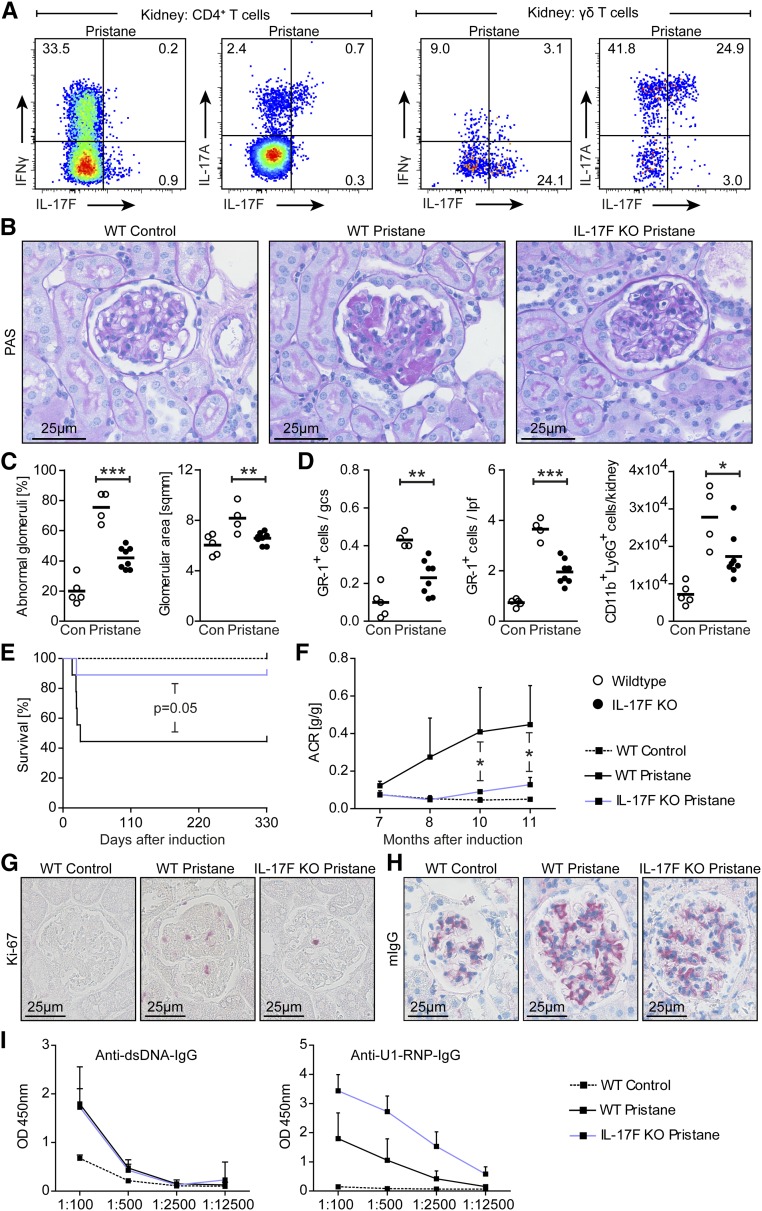
IL-17F promotes autoimmune disease in pristane-induced lupus nephritis. (A) Representative FACS plots showing IFNγ, IL-17F, and IL-17A expression after PMA/ionomycin stimulation by CD4+ T cells and γδ T cells isolated from kidneys of wild-type and IL-17F–deficient mice pregated for live CD45+ cells 11 months after disease induction. (B) Representative photographs of PAS-stained kidney sections from control mice, wild-type lupus mice, and IL-17F–deficient lupus mice 11 months after disease induction (original magnification ×400). (C) Quantification of glomerular abnormalities and glomerular area of controls (n=5), wild-type lupus mice (n=4), and IL-17F–deficient lupus mice (n=8) 11 months after disease induction. (D) Quantification of immunohistochemically stained tubulointerstitial and glomerular GR-1+ cells, as well as quantification of FACS analysis of renal neutrophils (defined as CD45+CD11b+Ly6G+ cells) in the aforementioned groups 11 months after disease induction. (E) Kaplan–Meier plot of survival in control and lupus nephritis groups over the course of disease. (F) ACR of the groups mentioned above over the course of disease. (G, H) Representative photographs of (G) Ki-67- and (H) mouse IgG-stained kidney sections of the aforementioned groups 11 months after disease induction. (I) ELISA analyses of circulating mouse autoantibodies against dsDNA and U1-RNP from sera of control and lupus nephritis mice 11 months after disease induction. Symbols represent mean values±SD connected by lines, or individual data points with the mean as horizontal line. *P<0.05; **P<0.01; ***P<0.001.

