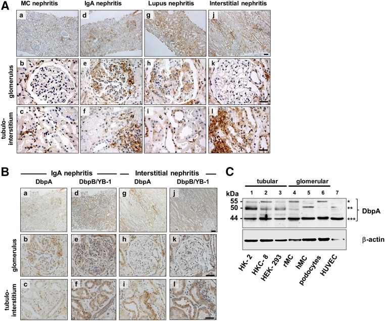
Figure 1.
Expression of DbpA in human kidney disease and cell lines. (A) Immunohistochemistry shows that DbpA expression is not detected in glomerular cells in minimal change (MC) GN, however DbpA expression is interspersed in tubulointerstitial cells (a–c). In contrast, DbpA protein was detected within the mesangial compartment of the glomeruli from patients with IgA nephritis and in cell infiltrates of the interstitium (d–f). A similar pattern of glomerular expression was observed in patients with lupus ISN/RPS class 4 G(A) GN with upregulated DbpA expression in tubular cells (g–i). The most profound tubular cell and interstitial upregulation of DbpA was seen with interstitial nephritis, where glomerular cells were immune negative (g–l). Images were made with a Leica DM6000 B Microscope (Leica Microsystems) using either a 100× (a, d, g, and j) or a 400× objective (b, c, e, f, h, i, k, and l). Scale bars, 200 μm in a, d, g, and j; 50 μm in b, c, e, f, h, i, k, and l. (B) Immunohistochemistry for DbpA and DbpB/YB-1 in sequential tissue slices from a patient with IgA nephritis reveals concordant upregulation of both proteins in mesangial compartment; at the same time, the staining pattern in the tubular cells and tubulointerstitial infiltrate markedly differs. (C) Western blot analysis of DbpA protein expression in cell lysates of human proximal tubular epithelial cells HK-2 and HKC-8, rMCs, human primary mesangial cells (hMCs), human embryonic kidney cells (HEK-293s), human podocytes, and human umbilical vein endothelial cells (HUVECs). Three major bands are shown: *55 kD; **50 kD; ***44 kD.

