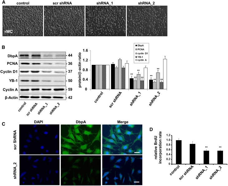
Figure 2.
DbpA knockdown leads to decreased cell proliferation. (A) Cellular morphology of rMCs after knockdown of DbpA for 3 days using two different shRNA oligos. (B) Western blot analysis confirms successful DbpA knockdown of transcripts by shRNA, which is accompanied by a significant reduction in the expression of cell proliferation markers (e.g., PCNA and cyclin D1) but not cyclin A. **P<0.01 (n=3). (C) Immunofluorescence staining observes both cytoplasm and nuclear expression of DbpA, but lower expression of DbpA protein in shRNA-incubated cells. Scale bar, 50 μm. (D) BrdU cell proliferation assay shows a marked decrease of cell proliferation in DbpA shRNA cells compared with the scrambled shRNA control. **P<0.01 (n=3).

