Figure 4.
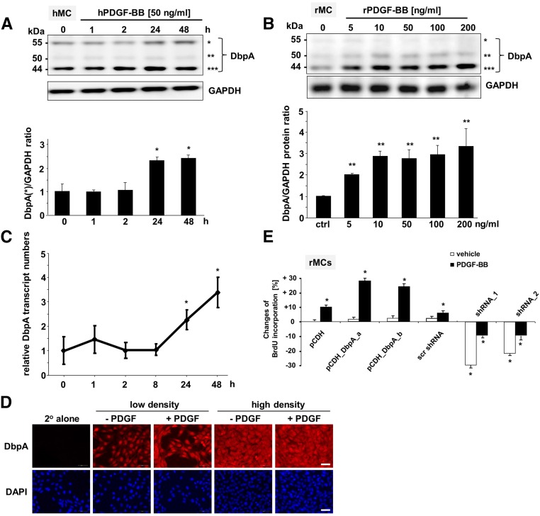
DbpA protein expression is upregulated by PDGF-BB stimulation in mesangial cells in vitro. (A, upper panel) Western blot analysis of DbpA protein expression in PDGF-BB–challenged primary human mesangial cells (hMCs). *55 kD; **50 kD; ***44 kD. (A, lower panel) Quantification of band intensities reveals 2.5-fold induction of DbpA protein expression (44 and 55 kD) after 24 hour incubation with human PDGF-BB (50 ng/ml). *P<0.05 (n=3). (B, upper panel) Western blot analysis of DbpA protein expression in rMCs stimulated with rat PDGF-BB for 24 hours at increasing doses. A dose-dependent upregulation of DbpA is shown from 5 to 10 ng/ml PDGF-BB stimulation. No significant change of DbpA expression was observed after stimulation with 50–200 ng/ml PDGF-BB. *55 kD; **50 kD; ***44 kD. (B, lower panel) Two bands at 50 and 44 kD are seen, with the predominant band at 44 kD. ctrl, Control. **P<0.01 (n=3). (C) Real–time PCR analysis for DbpA transcripts in rMCs reveals a profound upregulation after 24 and 48 hours of PDGF-BB incubation. *P<0.05 (n=3). (D) Immunofluorescence staining for DbpA in rMCs. Compared with vehicle, DbpA expression increases after 24 hours of incubation with rPDGF-BB (50 ng/ml). DbpA protein accumulates mainly within the cytoplasm. Scale bar, 50 μm. (E) Changes of BrdU incorporation rates are determined for rMCs undergoing overexpression of DbpA_a and DbpA_b or knockdown of DbpA. Stimulation with rPDGF-BB (50 ng/ml) was performed for 24 hours. *P<0.05 (n=3).

