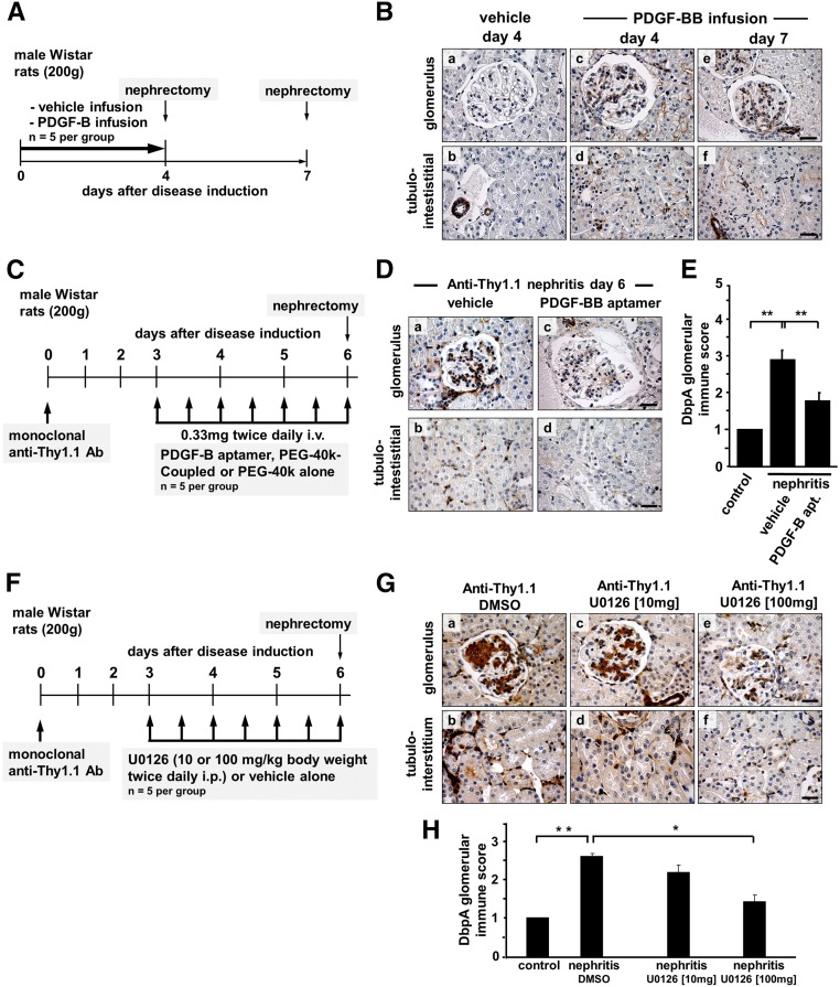
Figure 6.
Renal expression of DbpA after in vivo infusion of PDGF-BB. (A) Immunohistochemistry for DbpA was performed with renal biopsies from rats that were infused with vehicle for 7 days (control) or PDGF-BB for 4 or 7 days. (B) Whereas control rats express no DbpA in the glomeruli, the induction of DbpA is apparent within the mesangium of PDGF-BB–infused rats on both day 4 and day 7. In vivo treatment of anti–Thy1.1 nephritis rats with PDGF-B–specific aptamers leads to decreased DbpA upregulation. (C) After the induction of anti-Thy1.1 nephritis, animals received twice daily injections of PDGF-B aptamers or vehicle alone from day 3 to day 6 (schematically depicted). (D) Immunohistochemistry staining for DbpA reveals a remarkably decreased expression of DbpA in PDGF-B aptamer–treated rats (b and d) compared with vehicle-treated controls (a and c). Representative results are shown. Scale bars, 25 μm. (E) Quantification of the immune score of DbpA protein results in a significant decrease in anti-Thy1.1 nephritis treated with aptamers versus vehicle (P<0.01; n=5). In vivo treatment of anti–Thy1.1 nephritis rats with MEK inhibitor U0126 leads to decreased DbpA upregulation. **P<0.01. (F) After the induction of anti-Thy1.1 nephritis, animals received twice daily injections of U0126 (10 or 100 mg/kg) or vehicle alone from day 3 to day 6 (schematically depicted). (G) Immunohistochemistry staining for DbpA reveals a decreased expression of DbpA in U0126-treated rats (c–f) compared with vehicle-treated controls (a and b). Representative images are shown. Scale bars, 25 μm. (H) Quantification of the immune score of DbpA protein results in a significant decrease in anti-Thy1.1 nephritis treated with 100 mg/kg U0126 versus vehicle. *P<0.05 (n=5 for each group); **P<0.01 (n=5 for each group).

