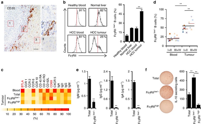
Figure 1. Accumulation of FcγRIIlow/− B cells correlated with disease progression and functional IL-10 production in HCC tumours.
(a) Representative distribution of CD20+ B cells in intratumoral (1), peritumoral stromal (2) and nontumoral (3) areas of HCC samples (n=10). Scale bar, 100 μm. (b) FACS analysis of FcγRII on CD19+ B cells from healthy blood (n=5), normal liver (n=2), and paired blood and HCC tumour (n=15). (c) Phenotypic characteristics of B cells, FcγRIIlow/− B cells or FcγRIIhigh B cells from paired blood and HCC tumour. Data for each of the indicated markers represent at least four patients. (d) Associations of tumour FcγRIIlow/− B cells with patients' TNM stage are shown (n=7 for stages I–II and n=7 for stages III–IV). (e) B cells (total) and sorted FcγRIIlow/− B cells from HCC tumours were stimulated with 1 μg ml−1 CD40L plus 50 ng ml−1 IL-21 for 6 days. IgM, IgG and IgA production were determined by ELISA. Results represent three independent experiments (n=5). (f) IL-10 production by total, FcγRIIlow/−, and FcγRIIhigh B cells from HCC tumours was determined by ELISpot (n=6). *P<0.05, **P<0.01 (Student's t test). Error bars, s.e.m.

