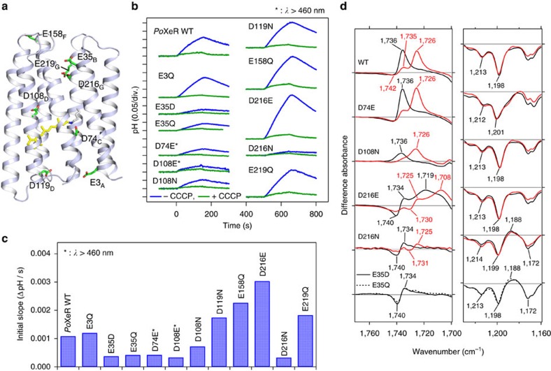
Figure 6. Inward H+ pumping activity and light-induced FTIR spectra of PoXeR and its mutants.
(a) The crystal structure of ASR (PDB ID: 1XIO)21 is shown with the side chains of the acidic residues used in PoXeR mutagenesis studies. The residue numbering of PoXeR is shown. (b) H+ transport activity of PoXeR mutants in E. coli cells after normalization for the amount of protein. Light is on between 0 and 150 s. (c) The initial slopes of light-induced pH changes shown in b. (d) Light-induced difference FTIR spectra of WT PoXeR and the mutants at T=230 K and pH 8.0. Spectra are measured in H2O (black) and D2O (red).

