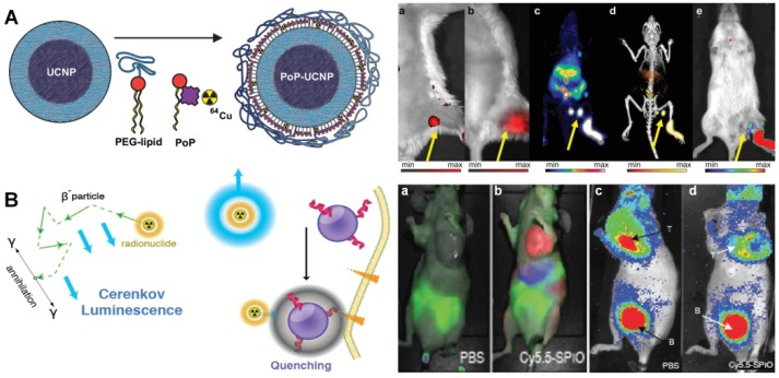
Figure 10.
(A) Schematic design of the PoP-UCNP structure. The animal imaging shows the mice injected with PoP-UCNPs and imaged in six modalities after 1 h injection, including (a) FL, (b) UC imaging. (c) PET, (d) PET/CT and (e) CL imaging, respectively. Reproduced with permission from reference 196. (B) Scheme of CL generation and nanoparticle induced quenching of CL. The animal imaging presents fluorescent imaging (a, b) and CR imaging (b, d) of mice injected with PBS and Cy5.5 labeled probe. B; bladder. Reproduced with permission from reference 203.

