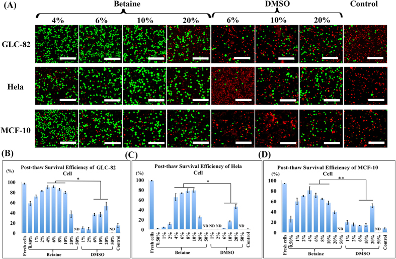
Figure 3. Cell cryopreservation using betaine with ultrarapid freezing.
(A) Fluorescence images of the live/dead assay of GLC-82 cells (upper row), Hela cells (middle row), and MCF-10 cells (lower row) cryopreservation with different concentrations of CPAs (betaine and DMSO), and cryopreservation in culture medium as control. Post-thaw survival efficiency of (B) GLC-82 cells, (C) Hela cells, and (D) MCF-10 cells evaluated at different concentrations of CPAs and control, with an identical cell amount (1.0 × 10⁶). Green: live cells. Red: dead cells. ND: not detected. Scale bar = 50 μm. Value = mean ± standard deviation, n ≥ 3. p < *0.05; **0.01.

