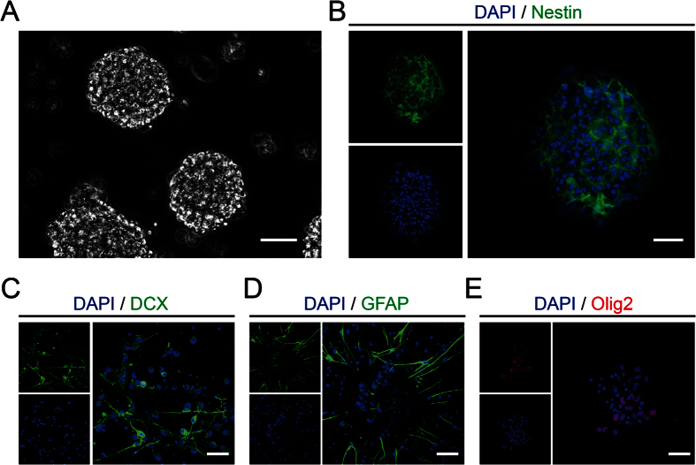
Figure 1. Characteristics and differentiation potential of NSPCs isolated from rats.
(A) The suspended growth of neurospheres was notably observed after 3 days. (B) The immunostaining showed the Nestin expression (green) on NSPCs in neurospheres before seeded on substrates. (C–E) The immunostaining demonstrated the differentiation potential of NSPCs into neurons (DCX, green), astrocytes (GFAP, green), or oligodendrocytes (Olig2, red). Cell nuclei were stained with DAPI in blue. Scale bar: 20 μm.

