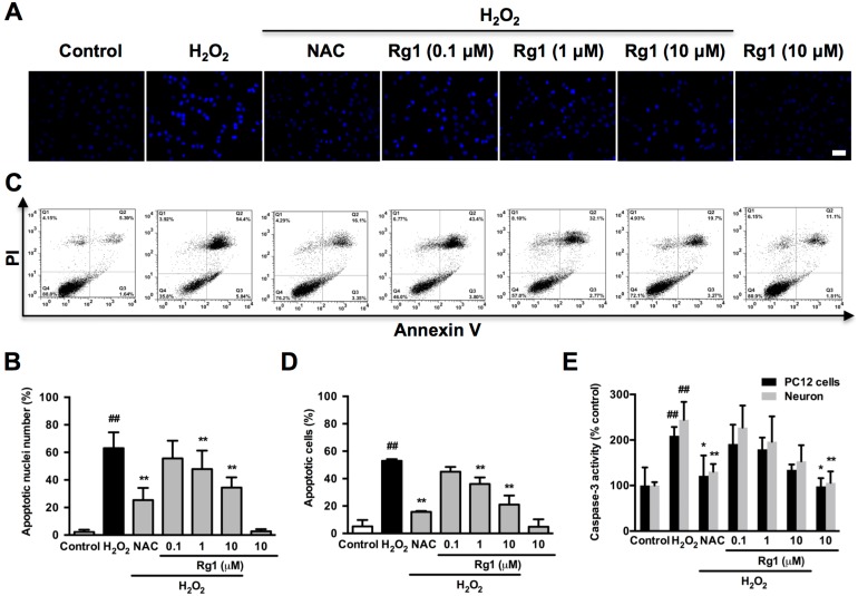
Figure 2.
Protective effects of Rg1 on H2O2-induced apoptosis in PC12 cells and neurons. PC12 cells and neurons were pre-treated with different concentrations of Rg1 (0.1, 1 and 10 μM) for 12 h prior to 100 μM H2O2 treatment for 12 h. NAC (500 µM) treated cells was tested as positive control. (A) Nuclear morphology by Hoechst 33342 staining in PC12 cells. Bar, 10 μm. (B) Quantification of condensed nuclei. (C) Flow cytometric analysis of Annexin V-FITC/PI-stained PC12 cells. Viable cells are Annexin V- and PI-, early apoptotic cells are Annexin V+ and PI- and late apoptotic cells are Annexin V+ and PI+. (D) The quantitative results were represented as the percentage of Annexin V-FITC+/PI+ cells among total cells. (E) Caspase-3 activity. Results were obtained from three independent experiments and were expressed as mean ± SD (##P< 0.01 versus control, *P< 0.05 versus H2O2-treated cells, **P< 0.01 versus H2O2-treated cells).

