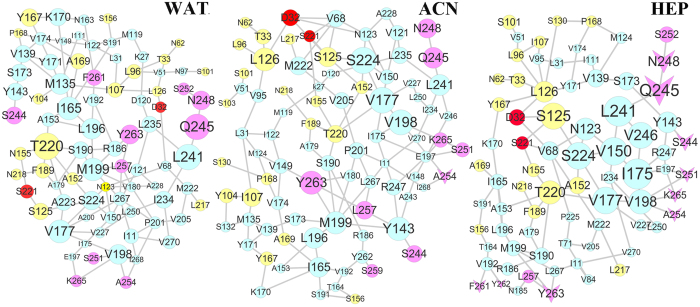
Figure 12. Protein structure networks (PSN) for the immobilized subtilisins in the three solvents.
Community networks from the adsorbed residues of SC (rose node) in the final snapshot of the 100 ns trajectories of CNT-wat system to the catalytic triad (red node) and the substrates binding pocket residues (yellow node) in aqueous (WAT), acetonitrile (ACN), and heptane (HEP) media, derived from the last 20-ns trajectories. The other residues in the pathways were colored as cyan. The residues desorbed in the organic media are displayed as V-shaped nodes. The size of the node is in proportion to the frequency of appearance in the pathways.

