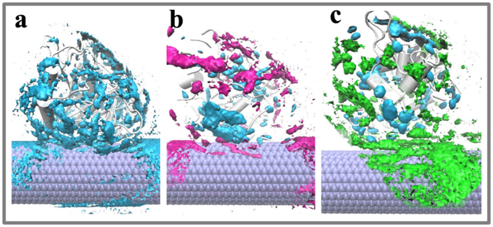
Figure 2. Spatial distribution of the solvent molecule around the immobilized enzyme.
(a) Aqueous solution (b) Acetonitrile media (c) Heptane media. The enzyme corresponds to the average structure of subtilisin over the last 20 ns equilibrium trajectory for each system. Solvent color code: water (cyan), acetonitrile (rose), heptane (green).

