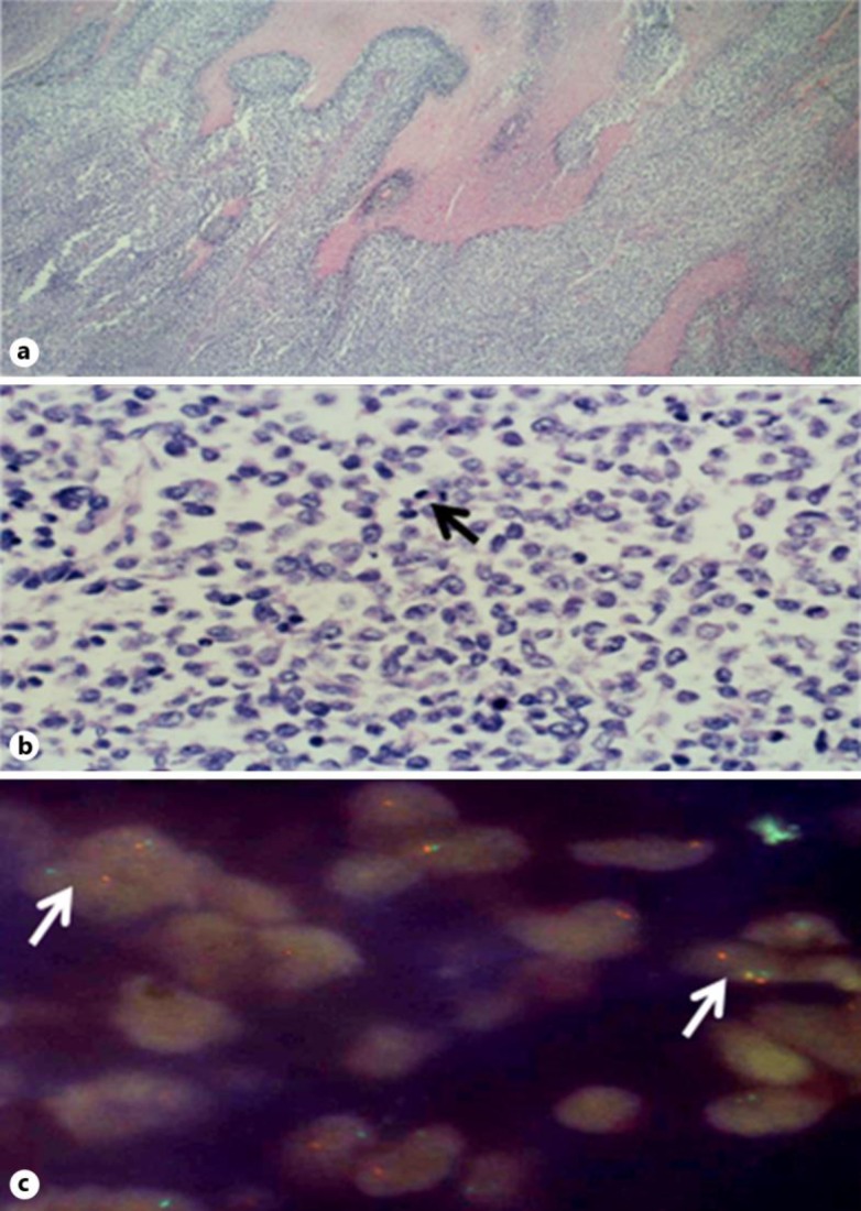
Fig. 1.
a Photomicrograph of the mass in the stomach wall showing a solid tumor with areas of necrosis. b Higher magnification shows cells of intermediate size, irregular nuclei, and clear cytoplasm with numerous mitoses (arrow). Immunohistochemistry stains for FLI1 and CD99 were positive. c FISH for 11: 22 translocation EWSR1 rearrangement was positive in 37% of cells. Positive cells show dissociation of green and orange probes (arrows).

