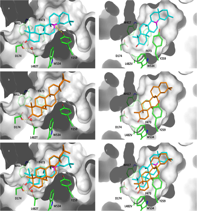
Figure 7. Modelled binding mode and poses overlays within MiFRS mutated.
(a) Modelled binding mode of oleanyl cation (cyan), (b) β-amyrin (orange) and (c) poses overlay within MiFRSLeu482Thr mutant binding site: the water molecule is indicated as red sphere, and polar interaction between Tyr259 and oleanyl cation (magenta) is indicated as yellow dashed lines. (d) Modelled binding mode of oleanyl cation (cyan), (e) friedelin (orange) and (f) poses overlay within MiFRSLeu482Val mutant binding site: polar interaction between Tyr259 and oleanyl cation (magenta) is indicated as yellow dashed lines.

