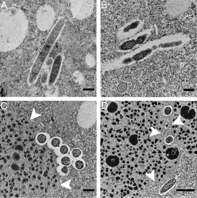
FIG 5.

Transmission electron microscopic images of “Candidatus Bealeia paramacronuclearis,” as observed for Paramecium biaurelia isolates US_Bl 11III1 and US_Bl 15I1. The symbiotic bacteria are oriented in parallel, in groups (A, B, and C), and were often found associated with the macronucleus (C and D), sometimes even lying in its folds (D). White arrowheads indicate the nuclear envelope. Scale bars, 0.5 μm (A, B, and C) or 1.0 μm (D).
