
 This work is licensed under a
 Figure 1.
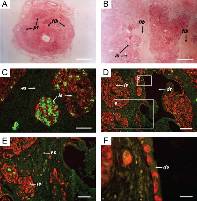
Photomicrographs of tissue sections from a biopsy of the patient’s pancreas. (A and B) Tissue sections stained with eosin, revealing the presence of diffuse fibrosis. (C and D) Immunostaining for insulin (green) and glucagon (red). Glucagon staining cells in islets, exocrine tissue and ductal epithelium. The boxed areas have been enlarged in panels E and F as indicated. (E) Further magnification of endocrine and exocrine pancreatic tissue, showing extra-islet glucagon staining cells (red). (F) Further magnification of ductal epithelium lined with cells staining for glucagon. Pt, pancreatic tissue; is, islet; ex, exocrine tissue; fib, fibrotic tissue; dt, duct; de, ductal epithelium. The bars represent 6 mm in A, 100 µm in B, C and D, 200 µm in E and 80 µm in F.