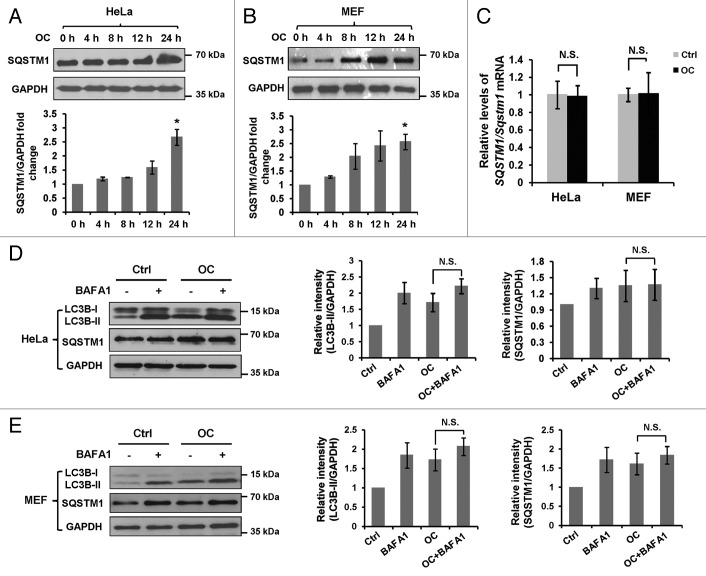
Figure 2. OC inhibits autophagic flux. (A) HeLa or (B) MEF cells were treated with OC (15 μM) over a certain time period (4, 8, 12 and 24 h), samples were analyzed by western blotting for endogenous SQSTM1 and GAPDH. ImageJ densitometric analysis of the SQSTM1/GAPDH ratio from immunoblots were shown (mean ± SD of 3 independent experiments, Student t test, *P < 0.05). (C) HeLa or MEF cells were treated with OC (15 μM) for 24 h. Relative SQSTM1/Sqstm1 mRNA levels (compared with GAPDH/Gapdh) was analyzed by quantitative RT-PCR. N.S., not significant. (D to E) HeLa or MEF cells were treated with DMSO or OC (15 μM) for 2 h in the presence or absence of 10 nM BAFA1 as indicated. Western blotting was performed to analyze the status of LC3B, SQSTM1 and GAPDH. ImageJ densitometric analysis of the LC3B-II/GAPDH and SQSTM1/GAPDH ratios from immunoblots is shown (mean ± SD of 3 independent experiments). N.S., not significant, Student t test.

An official website of the United States government
Here's how you know
Official websites use .gov
A
.gov website belongs to an official
government organization in the United States.
Secure .gov websites use HTTPS
A lock (
) or https:// means you've safely
connected to the .gov website. Share sensitive
information only on official, secure websites.
